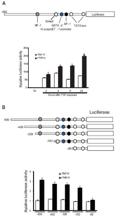
Figure 2. PreproET-1 promoter activity is stimulated by TNFα.
In the top portion of (A), the construct containing the −650 to + 170 region of the human preproET-1 promoter (PPET-1) that was utilized to determine the ability of TNFα to enhance ET-1 transcription (as in Methods) is shown. In the bottom portion of (A), luciferase activity of the preproET-1 promoter was tested at the indicted time points after exposure of stellate cells to TNFα (15 ng/mL) (n = 3; *p < 0.05 compared to control). The control (ctr) represents empty vector (pGL3) alone. In the top portion of (B), different preproET-1 promoter truncation constructs are shown. In the bottom portion of (B), luciferase activity observed 6 hours after TNFα exposure is shown (n = 3; *p < 0.05 compared to no TNFα).

