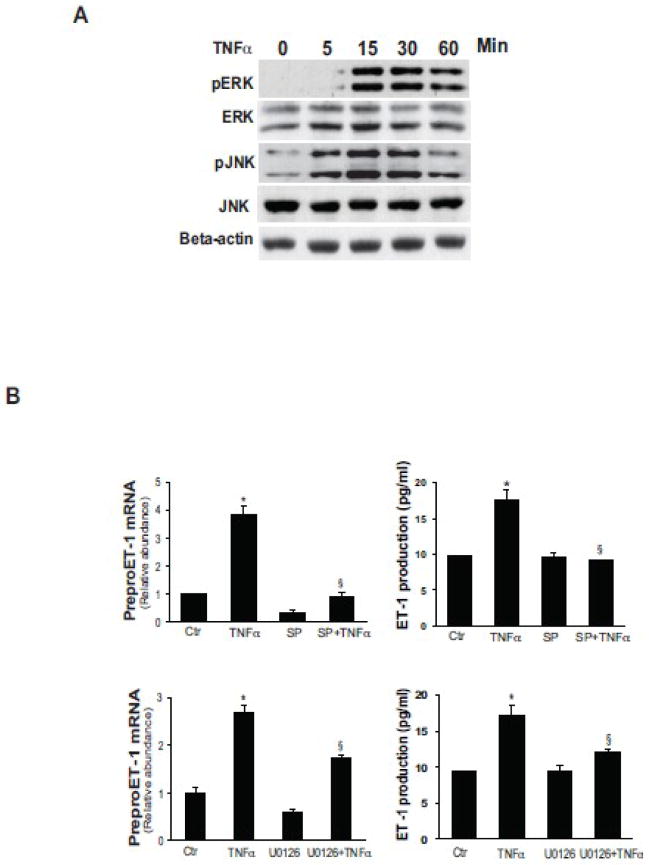
Figure 3. TNFα induces ET-1 expression through ERK and JNK.
In (A), activated stellate cells as in Figure 1 were exposed to TNFα (15 ng/mL) at the indicted times and ERK and JNK phosphorylation were detected by immunoblotting as in Methods. The blots shown are representative of 3 others. In (B), stellate cells as in (A) were exposed to TNFα (15 ng/mL) for 4 hours with the ERK inhibitor U0126 (10 μM, upper panel) or the JNK inhibitor, SP600125 (10 μM, lower panel), added 30 minutes prior to TNFα. PreproET-1 mRNA was detected by RT-qPCR (left panels) and immunoreactive ET-1 peptide (right panels) was measured as in Materials and Methods (n = 3, *p< .05 compared with control). Abbreviation: ctr = control; SP = SP600125

