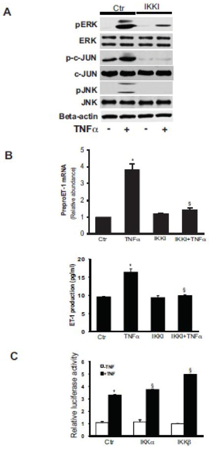
Figure 4. IKK mediates TNFα induced ET-1 synthesis.
In (A), activated stellate cells as in Figure 1 were exposed to TNFα (15 ng/mL) for 30 minutes with or without the IKK inhibitor (IKK Inhibitor 1, also IKKI, 200 nM), which was added 30 minutes prior to TNFα and whole-cell lysates were subjected to immunoblotting to detect the phosphorylated proteins as indicated as in Methods. The immunoblots shown are representative of 3 others. In (B), stellate cells were exposed to TNFα (15 ng/mL) for 4 hours with or without IKKI (200 nM). PreproET-1 mRNA (top graph) and immunoreactive ET-1 peptide (bottom graph) were detected as in Materials and Methods (n = 3, *p< 0.05 compared to control). In (C), IKKα and - β constructs were transduced into stellate cells and luciferase activity was measured, as in Methods (n = 3, *p< 0.05 compared to control; § p< 0.05 compared to TNFα control). Abbreviation: ctr = control.

