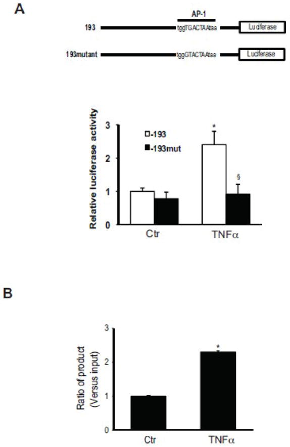
Figure 6. c-Jun activity regulates PPET-1 promoter activity and directly binds to the PPET-1 promoter region.
In the upper portion of (A), is shown a schematic diagram of mutated preproET-1 luciferase reporter vectors used to measure preproET-1 promoter activity. In the lower portion of (A), activated stellate cells as in Figure 1 were transfected with the above identified mutants, exposed to TNFα (15 ng/mL) for 6 hours minutes, and preproET-1 promoter activity was measured as in Methods. In (B), activated stellate cells as above were exposed to TNFα for 30 minutes, cells were harvested for ChIP assays. ChIP was performed with a specific antibody directed against phospho-c-Jun as in Methods. q-PCR was performed with primers for the AP-1 binding site (−108/−102) of the preproET-1 promoter; signals were normalized to those of input samples.

