
Figure 1. A&B. CD46 expression on NSCLC cells.
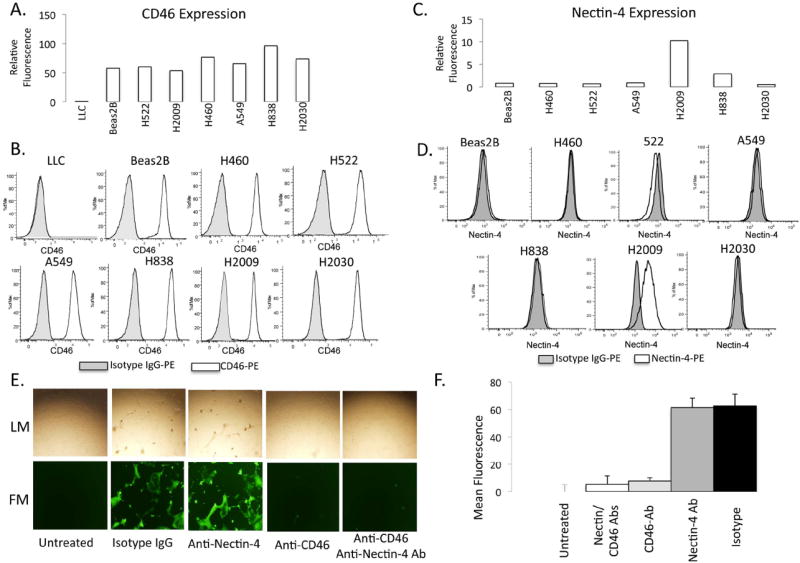
A) Plot of fluorescence intensity (relative to isotype) of cells stained with phycoerythrin-labeled anti-CD46 and assessed by flow cytometry. B) Representative histograms. LLC = murine Lewis Lung Carcinoma cell line as a negative control. C) Plot of fluorescence intensity (relative to isotype) of cells stained with phycoerythrin-labeled anti-Nectin-4 and assessed by flow cytometry. D) Representative histograms. E) Light (LM-top) and fluorescence (FM-bottom) microscopy of H2009 cells infected with MV-GFP after treatment with blocking antibodies against CD-46 and Nectin-4 or the combination. F) Quantification of fluorescence after blocking CD-46 and Nectin-4. Isotype IgG and H2009 cells untreated with MV-GFP were used as positive and negative controls, respectively.