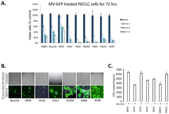
Figure 2. A-C. MV infects and lyses NSCLC cells in vitro.

A) Cell viability assay of cells treated with MV-GFP after 72 hours of treatment. Immortalized bronchial epithelial cells (HBEC and Beas2B) are used as a non-malignant control. (MOI = multiplicity of infection). Data are expressed as a percentage of untreated cells. B) Fluorescence microscopy of cells 72 hours following treatment with MV-GFP at MOI of 1.0 demonstrating fluorescent syncitia. Corresponding light microscopy photographs are shown. C) Human CEA measurement from supernatants of MV-infected NSCLC cells. Supernatants were collected 48 hours after infection of cells and measured by ELISA assay (CEA=carcinoembryonic antigen). Error bars indicate standard deviation of the mean.
