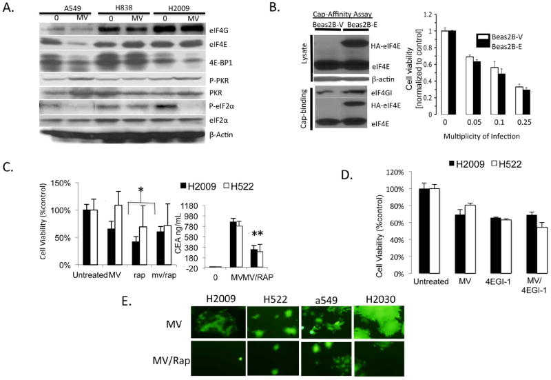
Figure 5. A-D. Effects of MV infection on protein translation signaling pathways.

A) H2009, A549, and H838 cell lysates were assayed by immunoblot before and 48 hours after infection with MV-GFP for key proteins involved in cap-dependent and independent translation initiation. B) Beas2B expressing empty vector (Beas2B-V) and Beas2B expressing eIF4E (Beas2B-E) were infected with MV-CEA at indicated MOI and assayed for cell viability 72 hours after infection. 5’ cap-affinity assay of untreated Beas2B-V and Beas2B-E are shown. Relative increases in eIF4G binding in the cap-affinity assay correspond to enhanced eIF4F cap-complex formation. C) H2009 and H522 NSCLC cells were treated with MV-CEA (MOI=0.01), rapamycin (Rap), or the combination and assayed for cell viability 72 hours after infection. Supernatants were collected from cells and assayed for CEA production as a measure of viral replication. Error bars indicate standard deviation of the mean. * indicates statistical significance. D) H2009 and H522 cells were treated with MV-CEA (MOI=0.01), 4EGI-1 (15μM), or the combination and cell viability assayed 72 hours after infection. E) NSCLC cell lines were treated with MV-GFP at MOI of 0.25 either alone or in combination with rapamycin. Representative fluorescence micrographs are shown.
