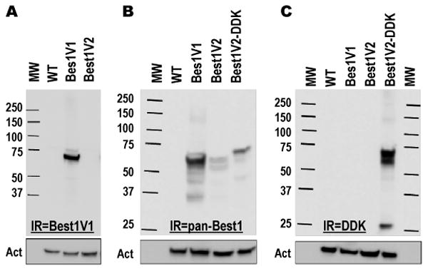
Figure 2. Western blot analysis of previously cloned splice variants of Best1 heteroexpressed in HEK293 cells.

A, Best1V1 and Best1V2 were heteroexpressed in HEK293 cells and probed with a monoclonal anti-Best1V1 antibody. B, Best1V1, Best1V2, and Best1V2 with a DDK epitope fused to the C-terminus (Best1V2-DDK) were heteroexpressed in HEK293 and probed with a polyclonal antibody recognizing Best1V1 and Best1V2. C, The same cell lysates as in B were probed with an anti-DDK antibody in order to assure specificity of the Best1V2 signal. For all Western blots the membranes were stripped and reprobed with a monoclonal anti-actin antibody to assess equality of loading (shown in boxes below). The figures are representative images of at least three Western blots performed in three independent cell transfections. Abbreviations: MW, molecular weight standards; WT, lysates prepared from mock-transfected (wild type) cells; Act, actin immunoreactivity. IR = immunoreactivity.
