
Figure 5. Biotinylation assay of the surface protein expression for three distinct splice variants of Best1 expressed in HEK293 cells.
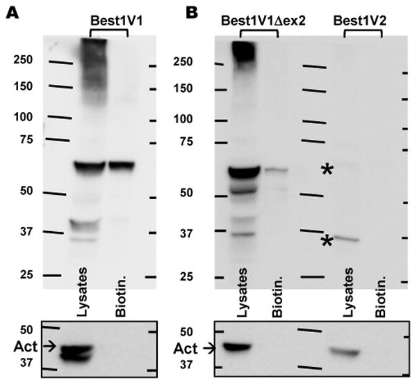
A, Western blot analysis of precipitated biotinylated fraction and the crude cell lysates in HEK293 cells expressing the DDK-tagged Best1V1. Representative of four blots. Because of the difference in the expression levels/intensity of the signal, Best1V1-DDK has been probed separately from Best1V1Δex2-DDK and Best1V2. B, Western blot analysis of biotinylated (surface) material and the crude cell lysates in HEK293 expressing Best1V1Δex2-DDK and Best1V2-DDK. Representative of three blots. Asterisks in (B) indicate two very weak immunoreactive bands in cells expressing Best1V2-DDK. Boxes below each panel show actin immunoreactivity on the same membrane after stripping. These controls show lack of cytosolic proteins in biotinylated fraction.