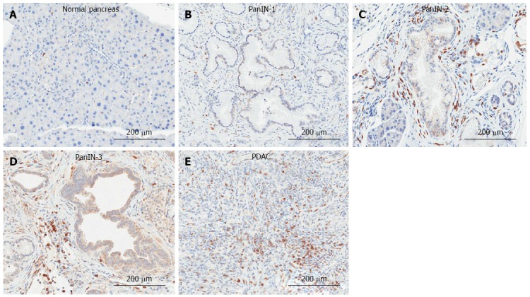
Figure 5.

Immunohistochemical detection of T-lymphocytes in the KrasG12D-induced mouse model of pancreatic ductal adenocarcinoma. A: Normal pancreas; B: Pancreatic intraepithelial neoplasia (PanIN)-1; C: PanIN-2; D: PanIN-3; E: Pancreatic ductal adenocarcinoma (PDAC) tissue was analyzed by immunohistochemistry for the presence of T-lymphocytes (CD3: abcam).
