. 2014 Aug 12;3:e26016. doi: 10.7554/eLife.03254

Figure 5. The identified cenRNA#1 transcript associated with CENP-A has a centromeric origin.

(A) CENP-A (green) was co-stained with Xist DNA FISH probe (FISH XistDNA, red) on chromatin fibers (cyan, DAPI). Two independent experiments were performed and in each, a minimum of eight chromatin fibers were analyzed per slide (co-localization on the same chromatin fiber between CENP-A and FISHXistDNA = 0/18). Scale bar: 1 µ m. (B) Chromatin fibers were either co-stained by IF/FISH with CENP-A (green) and cenRNA#1DNA (red), or IF/double-FISH with CENP-A (green), cenRNA#1DNA (red) and centromeric α-satellite probes (gray). The DAPI raw image is shown for a representative chromatin fiber (cyan). Two independent experiments were performed and in each, a minimum of eight chromatin fibers were analyzed per slide (co-localization on the same chromatin fiber between CENP-A and cenRNA#1DNA and FISH α-satellite compared to no co-localization = 9/20 vs 11/20). Scale bar: 1 µm. FISH, fluorescencein situ hybridization; IF, immunofluorescence.

Figure 5.

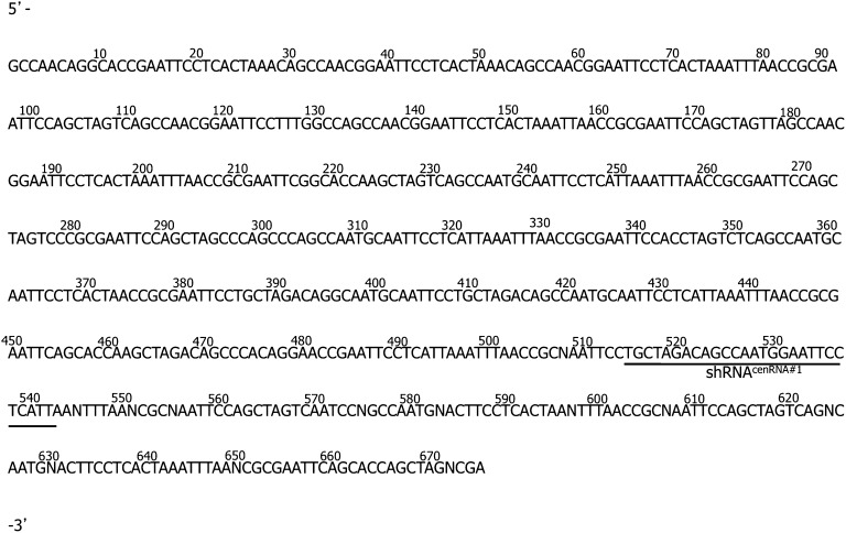
Figure 5—figure supplement 1. Sequence of cenRNA#1.

Figure 5—figure supplement 1.

RNAs immunoprecipitated with CENP-A were retro-transcribed and subcloned. One 675 nucleotide long sequence called cenRNA#1 was obtained by sequencing, and is reported here. The sequence of shRNAcenRNA#1 is underlined.
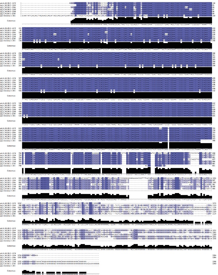
Figure 5—figure supplement 2. Alignment of the original cenRNA1 with new sequencing results.

Figure 5—figure supplement 2.

Adjustment was performed when similar uncalled bases (12 N’s) or called bases (27) were found in the six clones (~6% of total sequence changed).
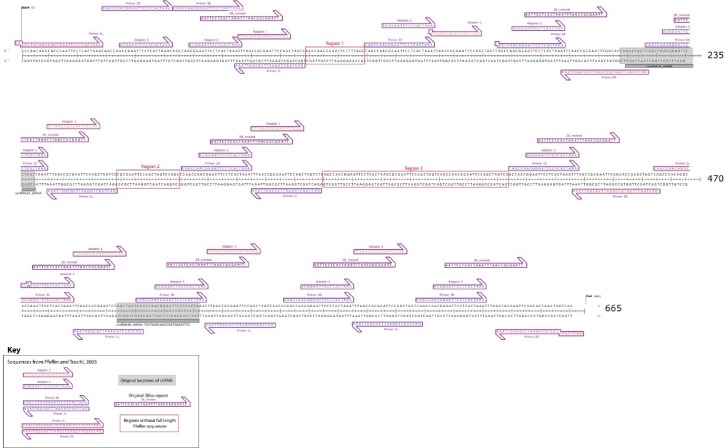
Figure 5—figure supplement 3. Map of re-sequenced cenRNA#1.

Figure 5—figure supplement 3.

Sequence alignments revealed that cenRNA#1 is principally composed of adapters used for cloning step described in (Pfeffer et al., 2005). Only full-length, 100% identical locations of Pfeffer et al. sequences are indicated (except at sequence ends).
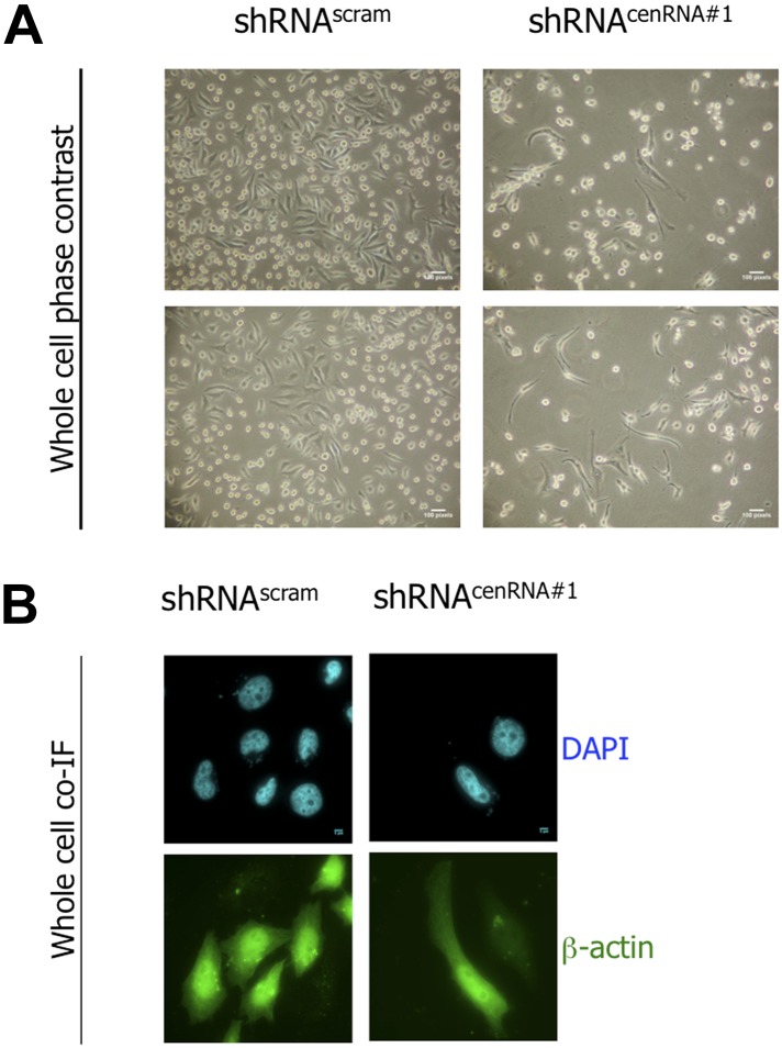
Figure 5—figure supplement 4. shRNAcenRNA#1-transfected cells have a cell survival defect. .

Figure 5—figure supplement 4.

(A) 6 days post-transfection of shRNAscram or shRNAcenRNA#1, cell morphology was observed by phase contrast microscopy. Scale bar: 100 pixels. (B) Cells were treated as in (A) and stained for β-actin (green) and DAPI (cyan) to reveal alterations in cell morphology. Scale bar: 5 μm. cDNA, complementary DNA; IF, immunofluorescence ; NT, non-treated.
Figure 5—figure supplement 5. The down-regulation of cenRNA#1 leads to chromosome defects. shRNAcenRNA#1-transfected cells have a cell survival defect. .

Figure 5—figure supplement 5.

(A) Transfected HeLa cells with cenRNA#1 was synchronized at mitosis - early G1. Then, cells were stained for CENP-C (marker of centromeres) and α-tubulin (marker of mitotic spindle). Mitoses were categorized for their chromosome defects (lagging chromosomes, chromosomal bridges or multi-polar spindles-MPS). Scale bar: 5 µm. (B) Mitotic defects were counted and percentages were diagrammed.