Figure 3.

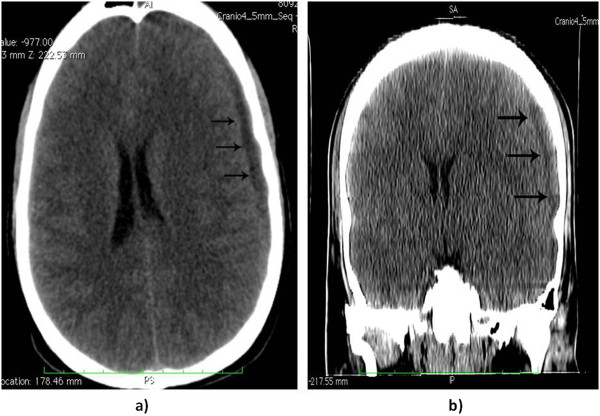
Computed tomography scan in axial (a) and coronal (b) cut: displaying the subdural collection (black arrows) of exudates on the left side in the temporal-parietal region. The air content of the empyema is noticeable.

Computed tomography scan in axial (a) and coronal (b) cut: displaying the subdural collection (black arrows) of exudates on the left side in the temporal-parietal region. The air content of the empyema is noticeable.