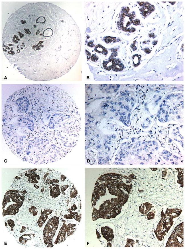
Figure 1.
WWOX immunohistochemical staining in normal and breast cancer tumor samples. (A) Representative low power photomicrograph from a TMA core of normal resting breast displaying strong staining in epithelial cells. (B) High power photomicrograph showing WWOX inmunostaining localizing to the cytoplasm of luminal epithelial cells. (C) TMA core representing IDCA completely negative for WWOX staining. (D) High power image of C. (E) TMA core representing IDCA displaying strong WWOX inmunostaining. (F) High power photomicrograph showing in detail strong cytoplasmic inmunostaining in nests and cords of epithelial malignant cells.

