
Figure 4.
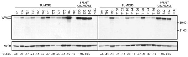
WWOX protein levels in normal and tumor breast tissue. Immunodetection of WWOX in human tissue extracts. Human breast protein extracts were separated by 12.5% SDS–PAGE and transferred to PVDF membranes. WWOX protein was detected using affinity purified rabbit polyclonal anti-WWOX antibody. The membrane was then probed with mouse monoclonal anti-actin antibody for normalization of differences in protein loading. Three normal human protein extracts prepared from breast organoids were included on each gel. WWOX protein levels in 23 breast tumors showed significant differences compared to normal breast WWOX levels. The full-length WWOX protein is indicated. NEG. – negative control extract (PEO1 cell line) that does not express WWOX protein. Rel. Exp.=Relative WWOX Expression. WWOX expression in each sample was first normalized to actin to correct for loading differences. WWOX expression in tumors relative to WWOX levels in normal organoids was then determined using the actin normalized values.