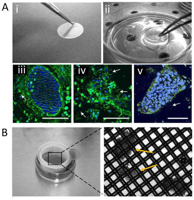
Figure 2.
(A) Surface-based vitrification method. (i) A modification of Thermanox cultivation disc with a small tip to handle with tweezer. (ii) Disc incubation in CPA. Multi photon laser scanning micrographs of hESC-colonies (iii) Control colony, (iv) Cryopreserved colony using slow rate freezing, and (v) Cryopreserved colony using surface based vitrification. Fewer membrane vesicles (arrows) were observed in vitrified sample. Scale bars indicate 100 μm. Reprinted by copyright permissions from [80]. (B) Bulk vitrification method. hESC cell clumps were loaded on the nylon mesh of a cell strainer and incubated in CPA. The inset (right) shows the magnified view of nylon mesh with cell clumps indicated by arrows. Reprinted by copyright permissions from [81].

