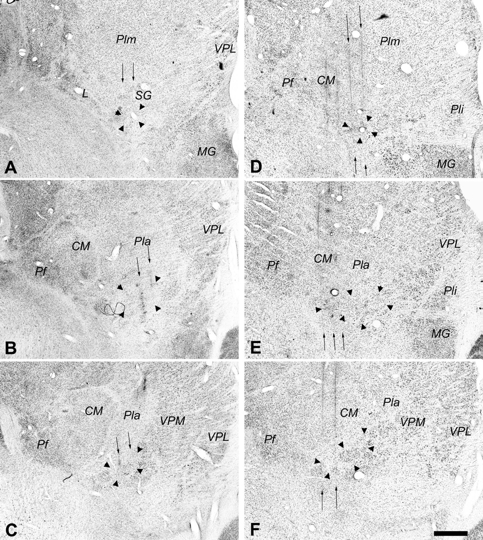
Figure 3.
Photomicrographs of coronal sections showing the histological traces of the microelectrode penetrations made in case m154 (left, panels A, B, C) and in case m57 (right, panels D, E, F). The borders of VMpo are indicated by large arrowheads. In each case, these photomicrographs show that penetrations were made through a very posterior level of VMpo (top panels, A, D), the middle of VMpo (middle panels, B, E), and a very anterior level of VMpo (bottom panels, C, F). The penetrations in which recordings were obtained from selectively nociceptive or thermoreceptive units and clusters are indicated by the thin vertical arrows, and the histology shows that these penetrations passed through VMpo. Bar = 0.5 mm. Abbreviations: CM, centre median; L, nucleus limitans; MG, medial geniculate; Pf, parafascicular nucleus; Pla, anterior pulvinar; Pli, inferior pulvinar; Plm, medial pulvinar; SG, suprageniculate nucleus; VPL, ventral posterior lateral nucleus; VPM, ventral posterior medial nucleus.

