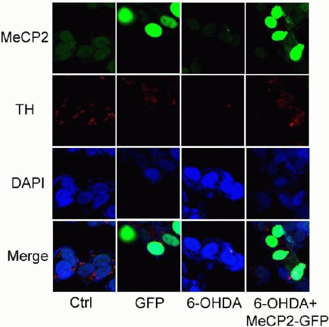
Figure 6.

Effects of upregulated X-linked methyl-CpG binding protein 2 (MeCP2) on 6-hydroxydopamine (6-OHDA)-treated SH-SY5Y cells (immunocytofluorescence staining, × 1 000).
Control (Ctrl): Untreated cells; GFP: cells transfected with pEGFP-N1 alone; 6-OHDA: cells incubated with 6-OHDA (50 μmol/L) for 24 hours; 6-OHDA + MeCP2-GFP: cells transfected with pEGFP-N1-MeCP2, followed by 6-OHDA (50 μmol/L) for 24 hours. Blue, green, and red fluorescence represent DAPI, MeCP2, and tyrosine hydroxylase (TH), respectively. After SH-SY5Y cells were incubated with 6-OHDA (50 μmol/L) for 24 hours, the red fluorescence became weaker, but after SH-SY5Y cells were transfected with pEGFP-N1-MeCP2, followed by 6-OHDA treatment for 24 hours, the red fluorescence did not become weaker.
