
Figure 2.
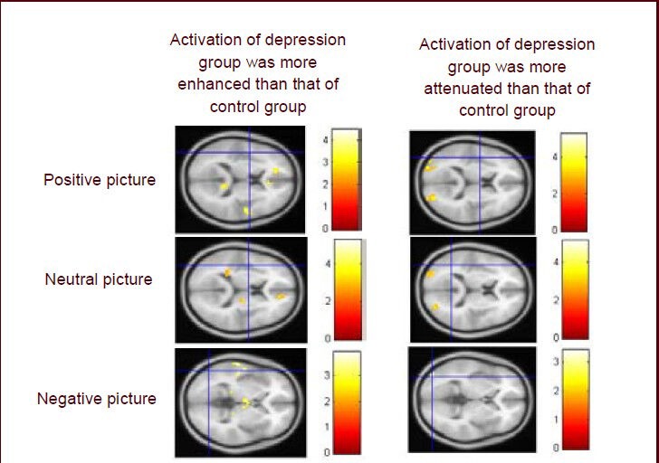
Functional MRI of activation of brain regions of patients with major depressive disorder.
The activation of some regions in the frontal lobe, temporal lobe, parietal lobe, limbic lobe and cerebellum was enhanced, but activation of some regions in the frontal lobe, parietal lobe and occipital lobe was weakened when the patients were watching positive and neutral pictures compared with normal controls. The activation of some regions in the frontal lobe, temporal lobe, parietal lobe, and limbic lobe was enhanced but activation of some regions in the occipital lobe was weakened when the patients were watching the negative pictures compared with normal controls. Colored bars reflect T scores for each analysis.