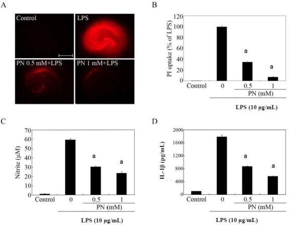
Figure 2.

Effects of paeonol on lipopolysaccharide (LPS)-induced hippocampal cell death.
Organotypic hippocampal slice cultures were pretreated with paeonol (PN) at the indicated concentrations for 30 minutes before adding 10 μg/mL LPS. After stimulation with LPS for 72 hours, the culture medium was collected and subjected to nitrite and cytokine assays, and was subsequently replaced with fresh serum-free medium containing propidium iodide (PI). The red color is the PI fluorescence, which indicates cell membrane damage. (A) PI fluorescence images. Scale bar: 500 μm. (B) Quantification of hippocampal cell death. Data are expressed as the percentage of the LPS value (the fluorescence intensity of the LPS group was designated as 100%). (C, D) Determination of nitrite (production of nitric oxide) and interleukin-1beta in culture supernatants. (B–D) The data were expressed as mean ± SEM from triplicate assays. Ten to 15 hippocampal slices were used in each group. aP < 0.001, vs. LPS-only treated group (Student's paired t-test). Organotypic hippocampal slice cultures incubated in the absence of LPS for 72 hours served as controls. M: mol/L.
