
Figure 4.
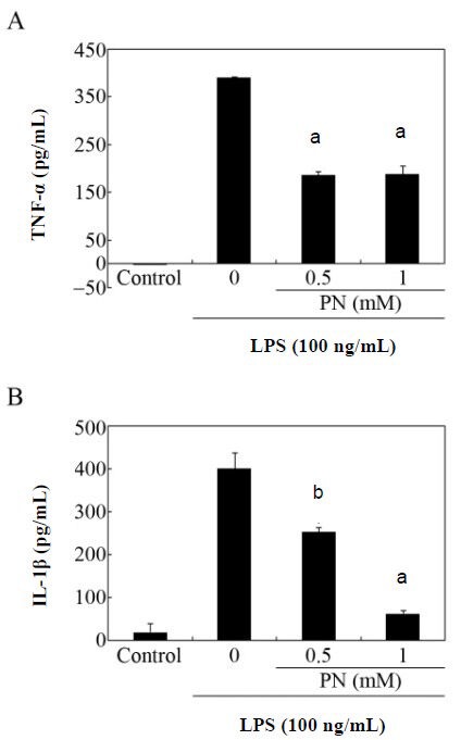
Effects of paeonol on the secretion of tumor necrosis factor-alpha (TNF-α) and interleukin-1 beta (IL-1β) by primary microglial cells.
Cultures were stimulated with lipopolysaccharide (LPS; 100 ng/mL) with or without pretreatment with the indicated amounts of paeonol (PN). After 24 hours of incubation, the culture supernatants were assayed by enzyme-linked immunosorbent assay (ELISA) for TNF-α (A) and IL-1β (B). Data are expressed as mean ± SEM from triplicate assays. aP < 0.001, bP < 0.05, vs. the LPS-only treated group (Student's paired t-test). The microglial cells incubated in the absence of LPS for 24 hours served as controls. M: mol/L.