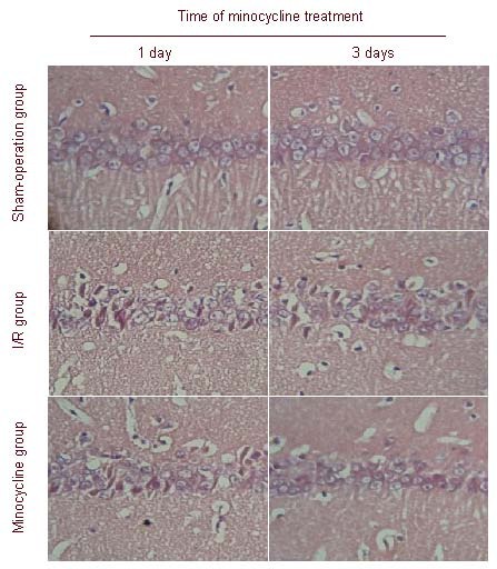
Figure 2.

Effect of minocycline on morphological changes in the hippocampal CA1 region of rats after cerebral ischemia-reperfusion (I/R) (hematoxylin-eosin staining, × 200).
In the sham-operation group, normal neurons arranged in neat rows, the cell bodies were full, and the outline of the nucleus was clear.
In the I/R group, neuronal necrosis in the hippocampal CA1 region and significantly less neurons were observed, the cell bodies were swollen and the number of pyramidal cells was decreased and lacked unity and coherence.
In the minocycline group, structural integrity of neurons increased significantly, and neurons slightly recovered in number.
