
Figure 6.
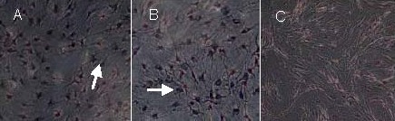
Morphology of glial fibrillary acidic protein-positive cells (arrows) after induction of bone marrow mesenchymal stem cells (immunohistochemical staining, × 200, streptavidin biotin-peroxidase complex method).
In the epidermal growth factor + basic fibroblast growth factor group (A) and epidermal growth factor + basic fibroblast growth factor + thrombospondin 1 group (B), glial fibrillary acidic protein-positive nuclei were stained dark blue, and the cytoplasm was stained as brown-yellow. The control group (C) showed no pigmentation.