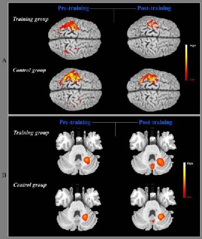
Figure 1.

Pre- and post-training changes in cerebral cortex activation during sequential finger movement using the right untrained hand in the training and control groups.
(A) Sequential finger movement induced cortical activation on the bilateral primary sensorimotor cortex, bilateral premotor cortex, and bilateral posterior parietal cortex in the first functional MRI scans in the training and control groups. Following 2 weeks of training with the left hand, activation on the ipsilateral premotor cortex and posterior parietal cortex decreased, and contralateral activation on the primary sensorimotor area, premotor cortex, and posterior parietal cortex decreased, in the training and control groups. However, the left primary sensory motor area activity was significantly decreased in the training group compared with the control group.
(B) In the first functional MRI scans for the cerebellar regions, the ipsilateral cerebellar areas (left dentate nucleus) showed activation during rapid sequential finger movement in the training and control groups, and greater activation of the medial cerebellar area (vermis) in the training group. After 2 weeks of training with the left hand, an obvious increase in neural activation was observed in the vermis and the left dentate nucleus. In the control group, cortical activation of the left dentate nucleus was slightly increased and the vermis was newly activated. In the highlighted bar for the activated peak voxel, the yellow color indicates higher neural activation than the red color.
