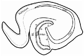
Figure 4.

Schematic drawing of the hippocampal subregions in a horizontal rat brain section, showing the hilus and molecular layer (ML) where ectopic granule cells (EGCs) were counted.
Granule cells in the area between the dotted line “a” and the outer border of the ML were counted as EGCs in the ML. The dotted line “a” is located three granule cell body widths from the outer border of the granule cell layer (GCL). The dotted line “b” indicates two granule cell body widths away from the inner border of the GCL whereas the dotted lines “c” and “d” connect the beginning of the CA3 pyramidal cell layer with the two ends of the GCL. Granule cells in the area enclosed by the dotted lines “b”, “c”, and “d” were counted as hilar EGCs.
