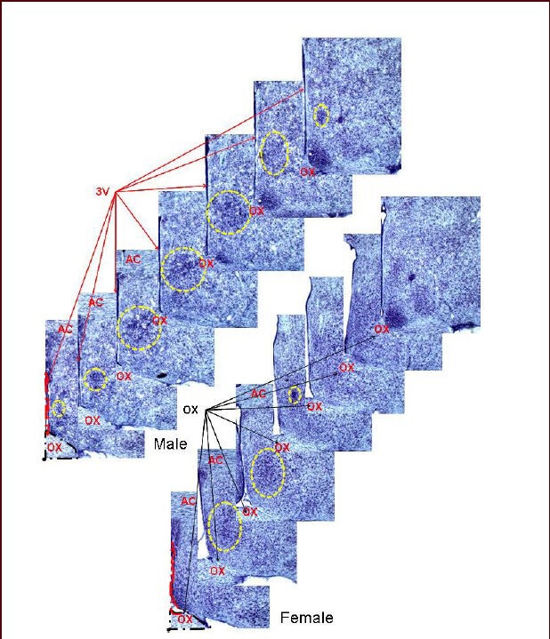
Figure 1.

Serial brain sections: the sexually dimorphic nucleus of the preoptic area stained with thionin.
Landmark structures that are also found throughout serial sections containing the sexually dimorphic nucleus of the preoptic area are labeled including the 3rd ventricle (3V) and the optic chiasm (OX). These structures were traced and their images used in constructing the three-dimensional images. The yellow dotted circle highlights the sexually dimorphic nucleus of the preoptic area. This figure is from our unpublished data (NCTR/US FDA Protocol P00710). Sexually dimorphic nucleus of the preoptic area: the area outlined by the yellow dashed circle; AC: anterior commissure. 10 × magnification was used.
