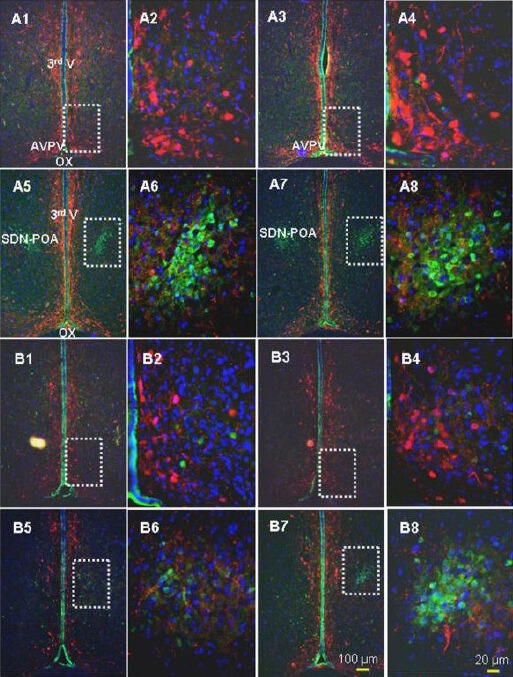
Figure 4.

Representative images of the triple labeling approach using calbindin D28k (calbindin-D28K; green), tyrosine hydroxylase (red), and 4’,6-diamidino-2-phenylindole (DAPI; blue).
Shown are four sequential slices along the longitudinal axis of the brain with intervals of 90 μm between adjacent slices. Here, the male AVPV (A1 and A3) and male sexually dimorphic nucleus of the preoptic area (A5 and A7) and the female AVPV (B1 and B3) and female sexually dimorphic nucleus of the preoptic area (B5 and B7) are outlined by dashed rectangles. The sexually dimorphic nucleus of the preoptic area is highlighted by calbindin-D28K immunoreactivity: no TH-positive cells were found, but fine axon-like projections/synaptic structures were seen. A neuron double-labeled with calbindin-D28K and TH was rare. The white rectangles in A1, 3 and B1, 3 highlight the AVPV, which is also displayed at higher magnification in A2, 4 and B2, 4. The white, dashed rectangles in A5, 7 and B5, 7 highlight the sexually dimorphic nucleus of the preoptic area, which is also displayed at higher magnification in A6 & 8 and B6 & 8. AVPV: Anteroventral periventricular nucleus; OX: optic chiasm; 3rd V: 3rd ventricle; SDN-POA: sexually dimorphic nucleus of the preoptic area. This figure is from our unpublished data (NCTR/US FDA Protocol P00706).
