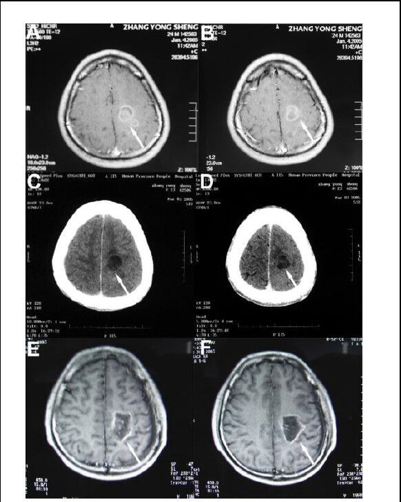
Figure 1.

A 24-year-old man with grade III recurrent glioma.
(A, B) Brain tumor showing an irregular enhancing signal shadow (arrows) on CT before treatment.
(C, D) CT scan showing internal liquefaction and tumor necrosis (arrows) in low-density images at 7 days after hyperthermia (hyperthermia + radiotherapy + chemotherapy).
(E, F) MRI scan showing increased tumor necrosis (arrows) and surrounding tissue with no obvious enhancement at 12 months after hyperthermia.
