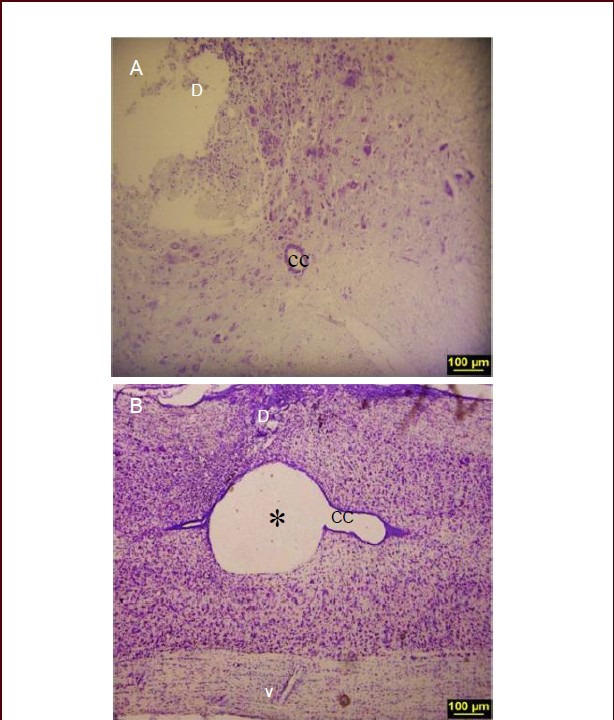
Figure 1.

Histological evidence of spinal cord lesion in spinal cord injury (SCI) rat model.
(A) Nissl-stained section illustrating the lesion in dorsal horn 1 week after SCI. (B) Nissl-stained longitudinal section at 3 weeks after SCI showing cavity formation (× 10). * indicates the cavity; D: dorsal; V: ventral, CC: central canal.
