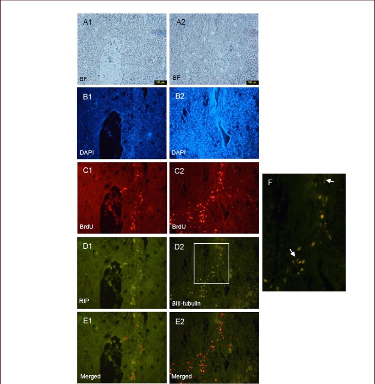
Figure 2.

An immunohistochemical procedure was used to detect receptor-interacting protein (RIP) and βIII-tubulin expression in rat spinal cord sections (× 10).
Images (A1, A2 bright field & B1, B2 DAPI) showing the cystic lesions forming at 3 weeks after spinal cord injury. The sections show that BrdU-labeled hair follicle stem cells survived for 3 weeks after transplantation, and the red cell aggregates (C1, C2) are clearly visible. The cells were assessed by double immunostaining. Through double label immunohistochemistry, the same section shows that cells were positive for the oligodendrocyte marker RIP (D1) and some of them were βIII-tubulin positive (D2). Also, cells with neuronal morphology are shown by arrows in image F. Moreover, BrdU-labeled cells that express both RIP and βIII-tubulin are shown in merged images (E1 and E2). D: Dorsal; V: ventral; BF: bright field; DAPI: 4’,6-diamidino-2-phenylindole; BrdU: 5-bromo-2’-deoxyuridine.
