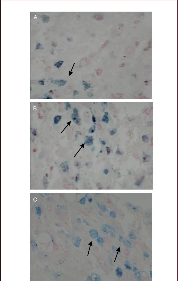
Figure 6.

Perl's Prussian blue staining of rabbit spinal cord injury tissue after transplantation of bone marrow mesenchymal stem cell-derived neuronal-like cells (phrase contrast microscope, × 200).
At 7 (A), 14 (B) and 21 days after cell transplantation (C). Arrows indicate cells containing blue-stained iron particles.
