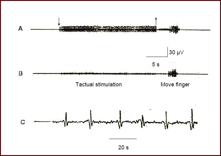
Figure 1.

The afferent nerve discharge of the superficial branch of the radial nerve.
The nerve action potential was detected using a guide electrode, and was input into a VC-10 storage phase oscilloscope and stored in a tape recorder.
(A) Nervous discharge (downward arrow represents stimulation onset, upward arrow represents stimulation end).
(B) Control record of the subcutaneous electrode located 1 cm lateral to the superficial branch of the radial nerve.
(C) Nervous system discharge recordings after amplification. The receptive field was the dorsal part of the second forefinger knuckle.
