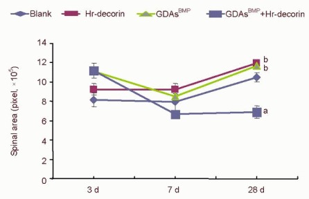
Figure 4.

Effect of combined GDAsBMP and hr-decorin transplantation on the cross-sectional area in the contused spinal cord of rats.
The cross-sectional area of the spinal cord in the glial fibrillary acidic protein-positive astrocyte fluorescence image was measured and analyzed with Image Pro Plus 6.0 software, and calculated by pixels. The cross-sectional area of combined GDAsBMP and hr-decorin transplantation group was least at 7 and 28 days (aP < 0.05, vs. the other groups at the same time); the cross-sectional area of hr-decorin group and GDAsBMP transplantation group was always larger than the blank group (bP < 0.05). Data are expressed as mean ± SD of six rats for each group (two-sample t-test and one-way analysis of variance).
Blank: Spinal cord injury group; Hr-decorin: human recombinant decorin injection group; GDAsBMP: glial-restricted precursor-derived astrocytes induced by bone morphogenetic protein-4 transplantation group; GDAsBMP + Hr-decorin: combined GDAsBMP and hr-decorin transplantation group.
