
Figure 1.
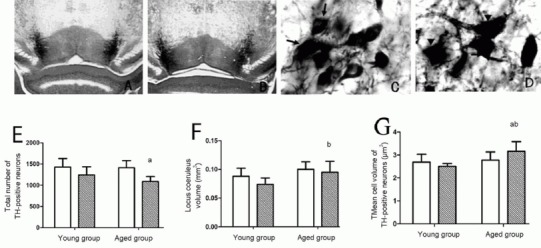
Pathological changes in locus coeruleus of wild-type and amyloid-β precursor protein and presenilin-1 double transgenic mice
(A, C) Tyrosine hydroxylase (TH) staining of noradrenergic neurons in the locus coeruleus of aged wild-type mice; (B, D) TH staining of noradrenergic neurons in the locus coeruleus of aged double transgenic mice. (E) Number of TH-positive neurons in unilateral locus coeruleus; (F) total volume of unilateral locus coeruleus; (G) mean volume of TH-positive cells in unilateral locus coeruleus.
Compared with wild-type mice, the total number of TH-positive neurons in the locus coeruleus (E) and total volume of the locus coeruleus were lower (F) in the aged groups, and the volume of cell bodies higher (D, G) in the aged groups. A, B: × 40; C, D: × 600; arrows: normal TH-positive cells; arrowheads: abnormal TH-positive cells. aP < 0.05, vs. aged wild-type group (aged group); bP < 0.05, vs. young double transgenic group (young group). Data are expressed as mean ± SD of six mice in each group (paired t-test). Black bars in E, F, G: double transgenic mice; white bar: wild-type mice.