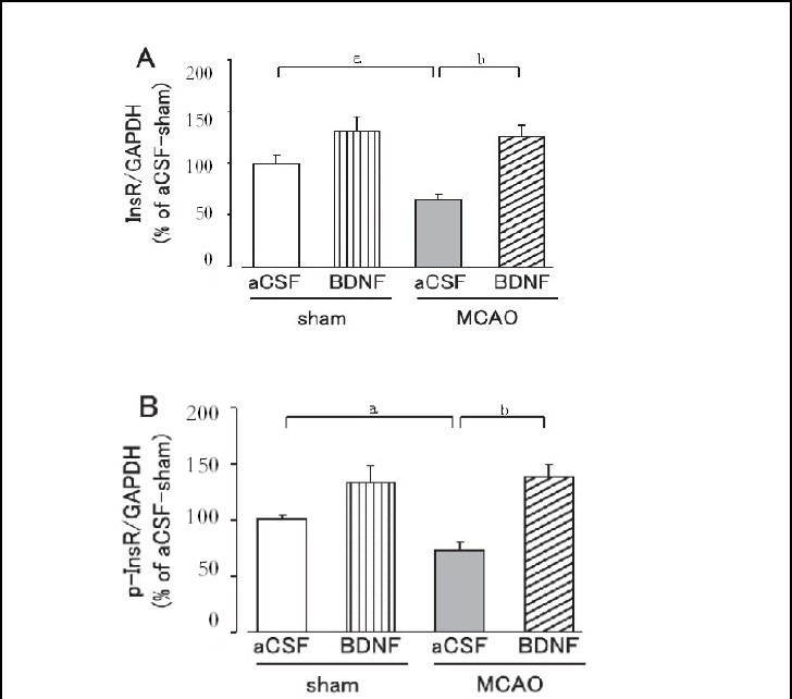
Figure 4.

Effect of brain-derived neurotrophic factor (BDNF) on hepatic insulin receptor (InsR; A) and tyrosine-phosphorylated insulin receptor (p-InsR; B) expression levels on day 1 after cerebral ischemia.
Mice were intrahypothalamically injected with BDNF (40 ng) or artificial cerebrospinal fluid (aCSF) immediately after middle cerebral artery occlusion (MCAO). The expression levels were detected by western blot analysis and analyzed by determining the absorbance ratio of InsR/ glyceraldehyde-3-phosphate dehydrogenase (GAPDH) (A) and p-InsR/GAPDH (B).
Data are expressed as mean ± SEM; n = 14 for the aCSF-treated sham group; n = 14 for the BDNF-treated sham group; n = 13 for the aCSF-treated MCAO group; n = 12 for the BDNF-treated MCAO group. aP < 0.05, vs. sham group; bP < 0.01, vs. aCSF-treated MCAO group using Student's t-test.
