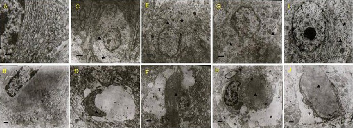
Figure 1.

Effects of olive leaf extract (OLE) on neuronal and capillary injury in the frontal lobe of the cerebral cortex in lead-poisoned mice (transmission electron microscope).
Ultrastructure of neurons (A, C, E, G, I, scale bars: 1 μm) and a blood capillary (B, D, F, H, J, scale bars: 0.5 μm) was observed.
(A, B) Normal group; (C, D) model group. In the model group, the neurons and capillaries showed severe injury, including nuclear membrane irregularities and rough endoplasmic reticulum dilation. The mitochondria were broken and vacuolated, the matrix around the capillaries appeared dissolved or destroyed, and the capillary lumen had narrowed.
(E, F) Low-dose OLE (250 mg/kg) group; (G, H) middle-dose OLE (500 mg/kg) group; (I, J) high-dose OLE (1 000 mg/kg) group. In these OLE groups, the ultrastructure of the cerebral cortex appeared much better, compared with the model group. The three doses of OLE showed protective effects on injured neurons and capillaries.
“*”: Nuclear membrane; “★”: rough endoplasmic retriculum; “▲”: mitochondrial vacaoles.
