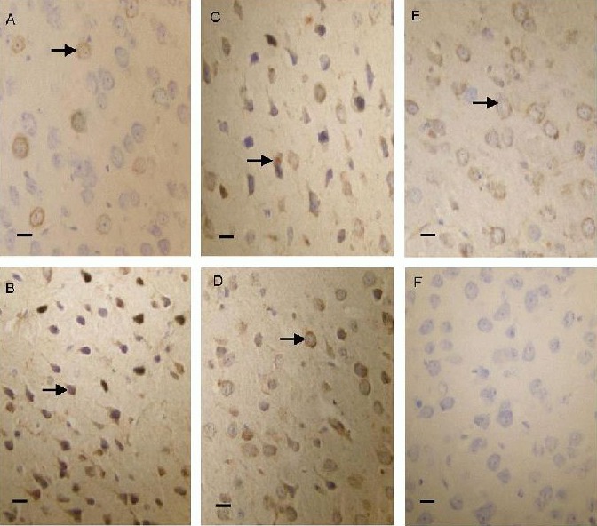
Figure 3.

Bax protein expression in the cerebral cortex of lead-poisoned mice (immunohistochemical staining, optical microscope, scale bars: 20 μm).
Bax protein was expressed predominantly in the cell membrane and cytoplasm. In the normal group (A), Bax-positive products were stained light yellow. In the low-dose olive leaf extract (250 mg/kg) group (C), middle-dose olive leaf extract (500 mg/kg) group (D) and high-dose olive leaf extract (1 000 mg/kg) group (E), the positive products were brown, and their levels were lower than in the model group (B). (F) Negative control group.
