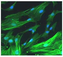
Figure 2.

Immunohistochemistry for myelin basic protein (green) of 4’,6-diamidino-2-phenylindole-labeled (blue) Schwann cells (fluorescence microscopy, × 200).
Purified Schwann cells were up to 95% or more pure, with a fusiform or constricted shape, and with small nuclei. Myelin basic protein immunofluorescence revealed the Schwann cell bodies.
