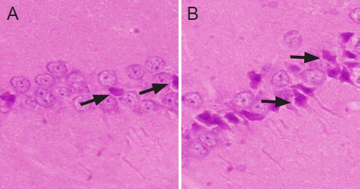
Figure 2.

Hematoxylin-eosin staining of rat hippocampus in the two groups (× 200).
(A) In the control group, only a small number of hippocampal pyra-midal cells were shrunken with condensed and deeply stained nuclear chromatin (arrows). (B) In the ischemia group, the majority of hippo-campal neurons exhibited pyknosis and deep staining (arrows). Some neurons and glial cells were significantly swollen and were round, and the space surrounding cells and capillaries was widened.
