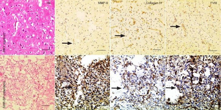
Figure 1.

Histological changes in brain tissues of stroke-prone spontaneously hypertensive rats with cerebral infarction.
Proliferative changes were visible in tissue. Cytoplasmic staining for matrix metalloproteinase-9 (MMP-9) is visible in vascular endothelial cells, astro-cytes, neurons, gitter cells and inflammatory cells. Moderate disruption of the microvessel basal lamina was revealed by staining for collagen IV. There was strong cytoplasmic staining for factor VIII (FVIII) in vascular endothelial cells in stroke-prone spontaneously hypertensive rats. Arrows show pos-itive expression. Scale bars: 50 μm. HE: Hematoxylin and eosin staining; WKY: Wistar-Kyoto; SHR-SP: stroke-prone spontaneously hypertensive.
