
Figure 3.
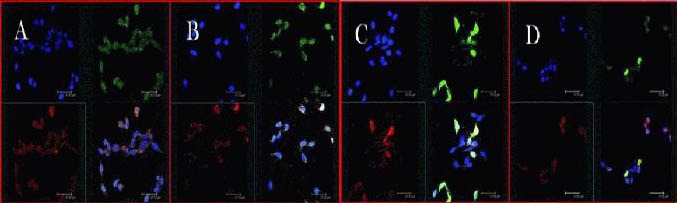
Confocal microscopic images of light chain 3 (red) and cathepsin B (green) immunohistochemistry at 1 day after middle cerebral artery occlusion.
Three kinds of staining were used in brain sections. Nuclei of neurons were labeled with blue, light chain 3 red and cathepsin B was labeled green. (A) Sham-surgery group; (B) model group; (C) rapamycin group; (D) 3-methyladenine (an autophagy inhibitor) group. Drugs were injected via the tail vein at 1 day after middle cerebral artery occlusion. The groups treated with 3-methyladenine showed an increased number of cells and enhanced cellular activities. Scale bars: 32.7 nm.