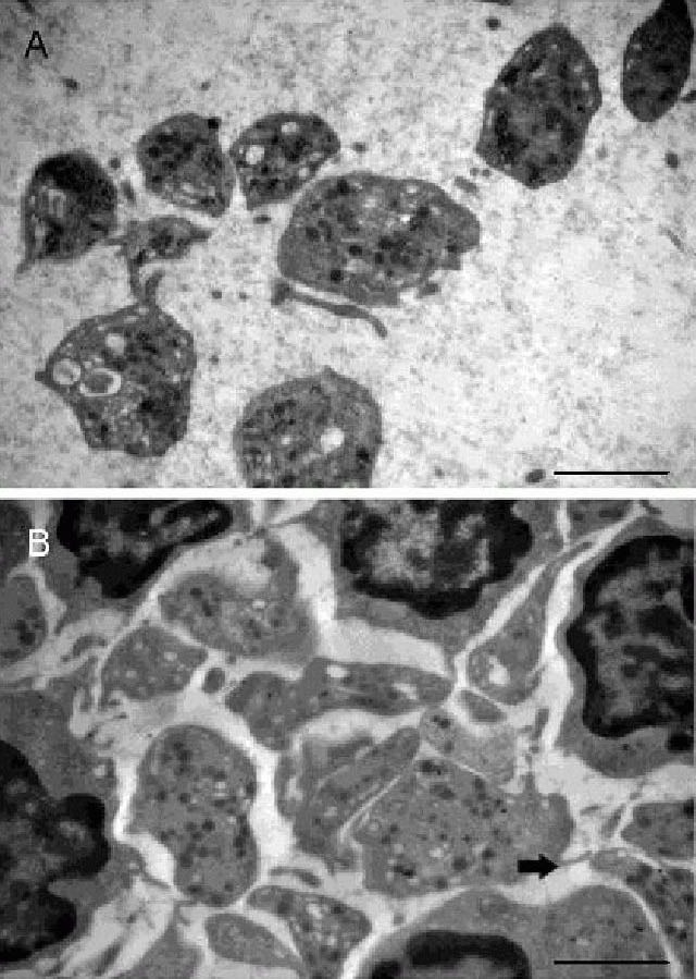
Figure 1.

Effects of acute cerebral infarction on platelet Golgi apparatus ultrastructure (transmission electron microscope, × 15 000).
Platelets from controls (A) showed clear profiles with more distributed alpha granules. Golgi tubules and vesicles were regularly arranged. In samples from 1, 7 and 15 days after infarction, aggregated platelets showed irregular morphology, more pseudopodium (arrow), and some platelet structures even disappeared. At each time point, alpha granules decreased visibly. Image from 1 day post infarction is used as a representation (B). Scale bars: 2 μm.
