Figure 1.

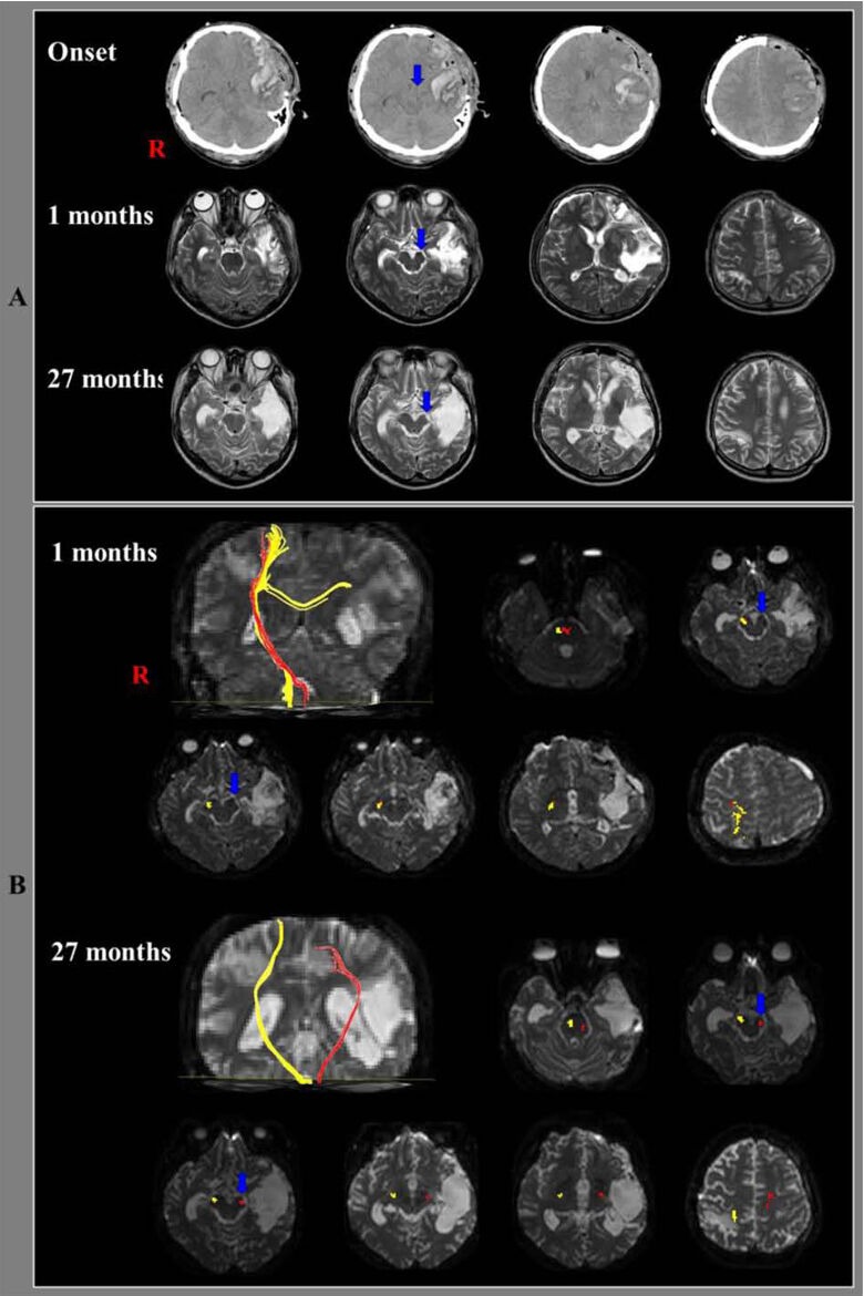
Brain CT, MRI images and diffusion tensor tractography results of the corticospinal tract in the included patient.
(A) Brain CT images after surgery show intracerebral hemorrhage in the left fronto-temporal lobes and left transtentorial herniation (arrow). Brain MRI images (1 and 27 months after onset) reveal shrinkage of the left cerebral peduncle (arrow). R: Right.
(B) Results of diffusion tensor tractography. The first (1 month after head trauma) and second (27 months after onset) diffusion tensor tractography for the corticospinal tracts (yellow) in the right hemispheres showed that fiber tracts passed along the known corticospinal tract pathway. On the first diffusion tensor tractography of the affected (left) hemisphere, the corticospinal tract (red) was disrupted below the cerebral peduncle (blue arrow) and connected to the right hemisphere via transpontine fibers. The transpontine connection fibers (red) in the right hemisphere may be related to compensatory mechanism after motor weakness or corticospinal tract injury. However, the left corticospinal tract (red) originated from the left primary motor cortex and descended through the left cerebral peduncle (blue arrow) on the second diffusion tensor tractography. R: Right.
