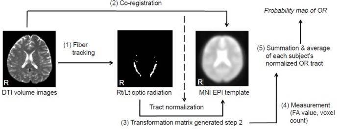
Figure 2.

The procedure for the quantitative measurement and probability pathway map reconstruction of the optic radiation (OR) fiber tract.
The OR fiber tracts from all subjects were extracted with diffusion tensor imaging (DTI) datasets, and brain normalization processes were per-formed in order to reduce individual variation and to allow for a comparison of the results under the same conditions without individual variation and to increase accuracy. The Montreal Neurologic Institute (MNI) echo-planar imaging (EPI) template was used to normalize the individual brains and extracted optic radiation fiber tracts. The volumetric analysis was performed with a voxel-count technique. The voxels through which the optic radiation fiber tract passed were counted and a percentage of their counted numbers according to whole-brain voxel numbers in the MNI EPI template was calculated. With the extracted optic radiation fiber tracts, the fractional anisotropy (FA) values were calculated, and a probability pathway map was acquired by averaging the normalized data for each subject for the OR fiber tract. Rt/Lt: Right/left; R: right.
