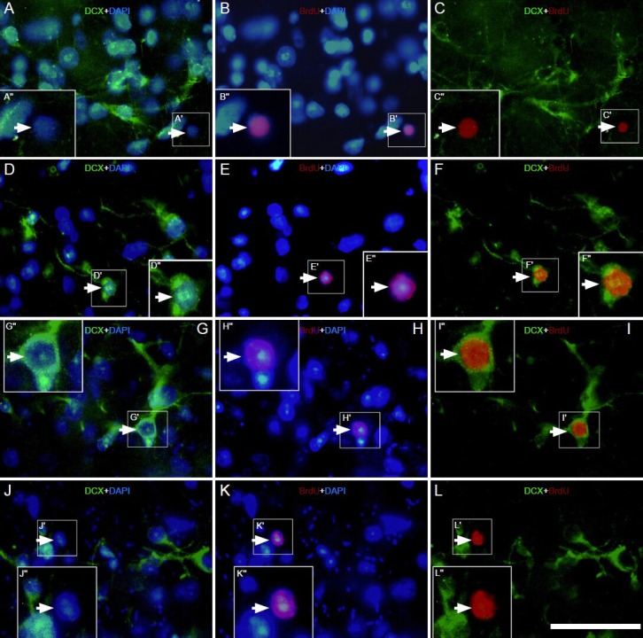
Figure 3.

Effect of environmental enrichment on bromodeoxyuridine (BrdU)/doublecortin (DCX) double-positive cells in layer II of the medial prefrontal cortex of guinea pigs (immunofluorescence staining).
Images immunostained with DCX (green), BrdU (red) and 4′,6-diamino-2-phenylindole (blue) showing the expression of BrdU/DCX double la-beled cells in the control group at (A–C) 0 days (D–F), 10 day (G–I) and 20 day (J–L) post-environmental enrichment. The big square insert (-”) is a high magnification view of the small frame (-′) in each image. The magnification of the objective lens was 20 × in A–L. The arrowheads indicate BrdU/DCX double labeled cells; the arrows indicate BrdU-positive cells without DCX colocalization. Samples were visualized via fluorescent mi-croscopy (Nikon, Eclipse 80i). Scale bar = 25 μm.
