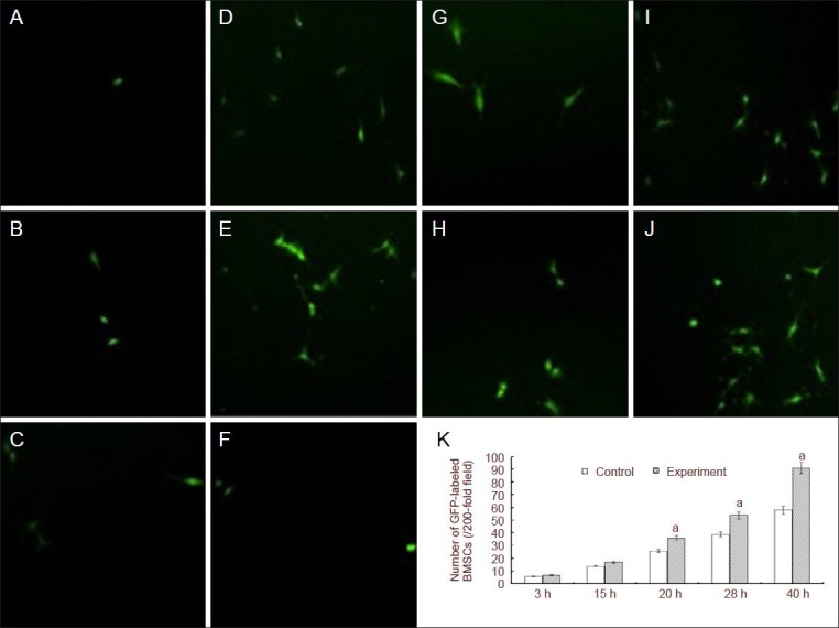
Figure 1.

Effect of red light irradiation on bone marrow mesenchymal stem cells (BMSCs) migrating towards primary neurons after oxygen-glucose deprivation (OGD).
(A–J) Green fluorescent protein (GFP)-labeled BMSCs in OGD control and phototherapy + BMSC groups at different culture times (fluorescence microscope, × 200). After 3 hours of culture, GFP-labeled BMSCs were visible in Transwell chambers and the number of migrated cells gradually in-creased as culture time proceeded. (A–E) 3-, 15-, 20-, 28-, 40-hour OGD control groups. (F–J) Phototherapy + 3-, 15-, 20-, 28-, 40-hour OGD groups. (K) Changes of the number of GFP-labeled BMSCs in OGD control and phototherapy + OGD groups at different culture times; h: hours. Data are expressed as mean ± SD. Differences between groups were compared using one-way analysis of variance, and pairwise comparisons were performed using Student-Newman-Keuls test. aP < 0.05, vs. control group. GFP gene transfection enables labeling of rat BMSCs with green fluorescence.
