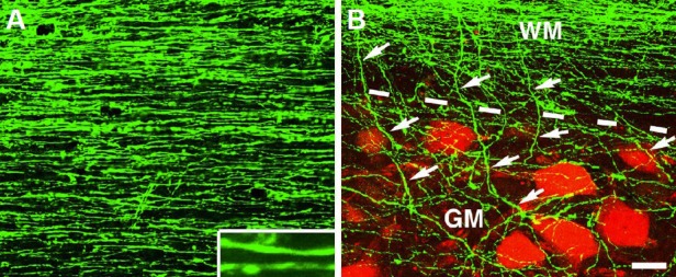
Figure 2.

Growth and innervation of human axons after spinal cord injury in rats.
(A) Growth of green fluorescent protein (GFP) expressing human axons (H9 derived) in the host white matter 3 mm caudal to a neural stem cell (NSC) graft placed in C5 hemisection site for 3 months. Inset shows individual axons at higher magnification in a horizontal section. (B) Innervation of human axons (green) from host white matter (WM) into gray matter (GM) is indicated by arrows. Host neurons are labeled with NeuN (red). Dashed lines indicate white matter and gray matter interface. Scale bar: 32 μm.
