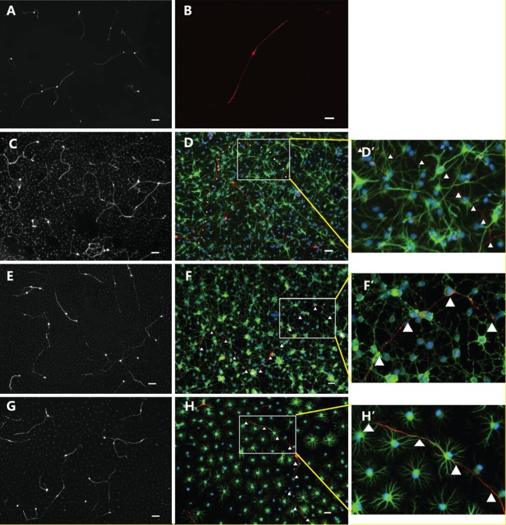
Figure 3.

Immunofluorescence images of dorsal root ganglion neurons co-cultured with various cells for 18 hours.
Under the fluorescence microscope, cells in the type-1 and type-2 astrocyte groups were stained with NF-M, glial fibrillary acidic protein (GFAP) and Hoechst 3442. Cells in the oligodendrocyte precursor cell group were stained with NF-M, O4 and Hoechst 3442. Cells in the blank control group were stained with NF-M and Hoechst 3442. (A, C, E, G) Low-power images of cells in the blank control, type-1 astrocyte, oligodendrocyte precursor cell and type-2 astrocyte groups were co-cultured with dorsal root ganglion neurons for 18 hours (B, D, F, H respectively). High-power images of cells in the blank control, type-1 astrocyte, oligodendrocyte precursor cell and type-2 astrocyte groups were co-cultured with dorsal root ganglion neurons for 18 hours. D’, F’, H’: Magnified images of white panes of D, F, H, respectively; red fluorescence shows NF-M staining. Green fluorescence in F and F’ shows O4 staining. In D, D’, H, H’, green fluorescence shows GFAP staining, and blue fluorescence shows Hoechst 3442-labeled nuclei. Scale bars (A, C, E, G): 100 μm; scale bars (B, D, F, H): 50 μm.
