Figure 3.

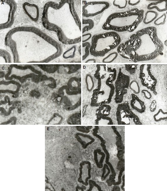
Effect of percutaneous microballoon compression on the ultrastructure of the trigeminal root at 7 and 14 days (d) after percutaneous microballoon compression (transmission electron microscope, × 3,000).
(A) In the normal group, myelin was arranged as homocentric circles and the stacking of the sheath was normal.
(B) In the 2-minute compression group at 7 days, large-diameter myelinated nerve axons were swollen, disrupted and demyelinated. Fragmentation of myelin and formation of digestion chambers were evident.
(C) In the 5-minute compression group at 7 days, part of the myelin had completely disintegrated into fragments. Significant demyelination was apparent.
(D) In the 2-minute compression group at 14 days, large-diameter myelinated nerve axons were strongly demyelinated and deformed. Large digestion chambers were evident.
(E) In the 5-minute compression group at 14 days, the myelin fragments were partially resorbed. Deformation of the large-diameter axons was apparent.
