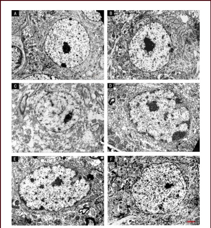
Figure 6.

Effect of scutellaria baicalensis stem-leaf total flavonoid (SSTF) on the ultrastructure of hippocampal neurons in cerebral ischemia-reperfusion injured rats (transmission electron microscopy, scale bar: 2 μm).
(A) In the control group, hippocampal neurons had clear boundaries and a large, central nucleolus, the plasma membrane was continuous and clear, and many organelles were visible. (B) In the sham group, the neuronal nucleolus was large, chromatin was uniformly distributed, the rough endoplasmic reticulum and ribosomes were abundant, and the Golgi and mitochondria were visible.
(C) In the model group, the majority of neurons were disintegrating, the cell membrane and nuclear membrane have ruptured, and nucleoli appeared significantly distorted. The chromatin condensed or dissolved, and the mitochondria had disintegrated. (D) In the SSTF low-dose group, the nuclear membrane and nucleolus were irregular, the cytoplasm appeared dissolved, and mitochondrial swelling or vacuolization was observed. The membranes of the endoplasmic reticulum and the Golgi were balloon-dilated.
(E) In the SSTF medium-dose group, hippocampal neurons were mildly swollen, the plasma and nuclear membrane were continuous, most of the mitochondria were intact, rough endoplasmic reticulum and ribosomes were abundant, and the Golgi was complete and clear. (F) In the SSTF high-dose group, hippocampal neuronal structures were normal, nuclear membrane was intact and the nucleolus was close to the center. Chromatin was uniform, mitochondria were intact, and the rough endoplasmic reticulum and ribosomes were abundant.
