
Figure 1.
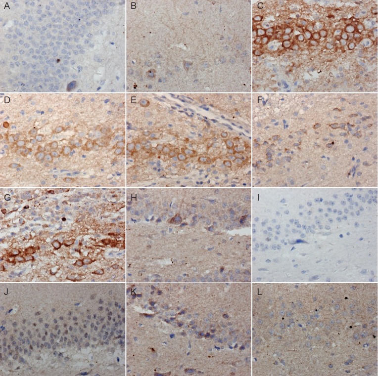
Immunohistochemical staining of axin (A–F) and β-catenin (G–L) in the hippocampal dentate gyrus of Alzheimer's disease rats with or without pretreatment with acupuncture and/or moxibustion (× 400).
Normal group (A: axin; G: β-catenin) and sham surgery group (B: axin; H: β-catenin) show few axin-positive cells but extensive β-catenin-posi-tive staining. Model group shows many axin-positive cells (C) but few β-catenin-positive cells (I). Electroacupuncture (D, J), moxibustion (E, K) and electroacupuncture + moxibustion (F, L) groups show similar numbers of axin- and β-catenin-positive cells to the normal and sham surgery groups.